
What is already known on this topic
Human microbiota-associated animal models relying on germ-free recipient mice are used to study the relationship between intestinal microbiota and human disease. However, transfer of microbiota into germ-free animals have raised concerns and limitations of this model system.What this research adds
In this study the researchers studied the colonization of the human and mouse microbiota transplanted to germ-free mice. Furthermore, the immune response in the recipient mice following transplantation has been characterized.Conclusion
This study revealed that immune-regulating bacteria are lost when transplanting microbiota from humans to laboratory mice and that the established human microbiota results in a weak stimulation of the murine immune system.
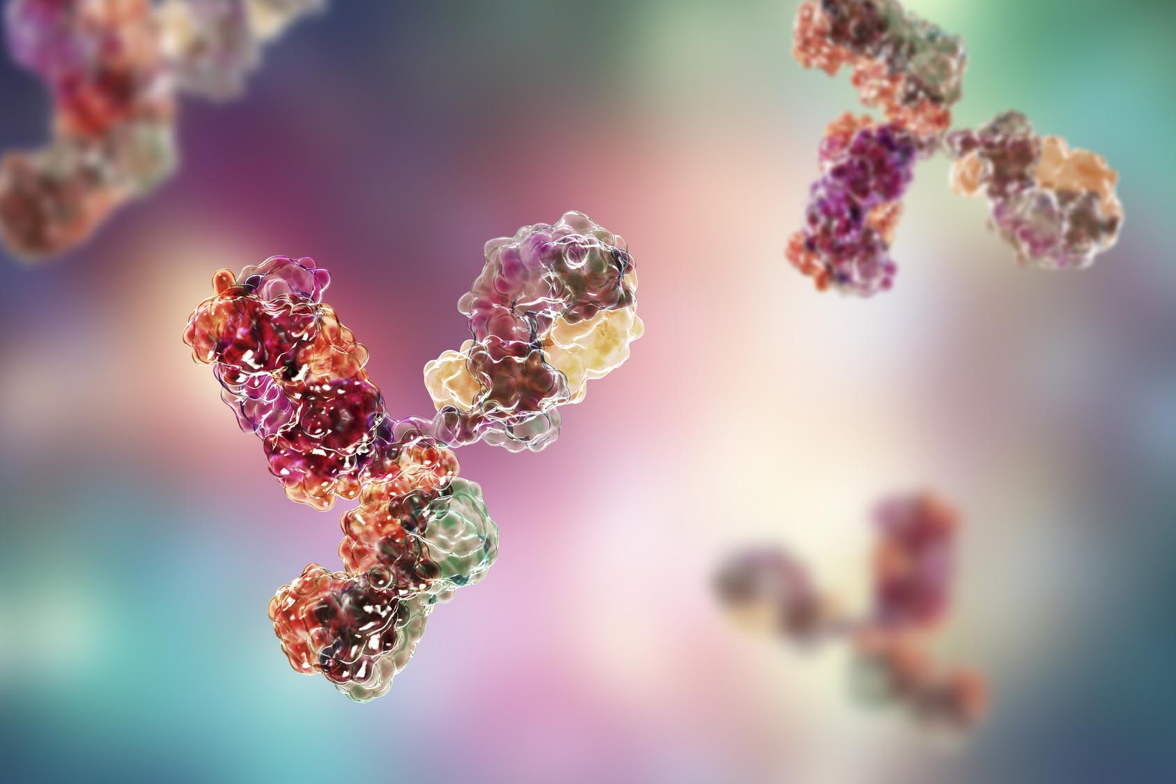
Microbiota transplantation is a treatment method that is gaining growing attention. It consists in transferring functional bacteria from one specie to another to remodel the intestinal flora structure and immune system, thereby achieving the purpose of treating diseases influence by microbiota. For years, mice have been transplanted with human microbiota (HM) although xenogeneic transplantation has shown some limitations probably due to immunological differences between donor and recipient.
Laboratory rodents are routinely raised in specific pathogen free (SPF) barrier facilities and while human bacteria are known to colonize the mouse gut better than environmental bacteria, has been shown that transplantation could trigger global changes in the recipient intestine, which can mask disease-specific attributes of the donor material. Therefore, more information on how to build an efficient model to replace microbiota of a developmentally mature intestinal environment remain highly desirable.
Currently, thanks to advanced sequencing techniques, it is possible to better identify and characterize the microbiome. In this study, the team used ribosomal RNA amplicon sequencing and taxonomy to investigate on how the intestinal immune system of germ-free (GF) mice is affected by transplantation with microbiota from mice or humans.
The researchers have transplanted germ-free (GF) C57BL/6 (B6) inbred mice with a xenogeneic HM. Also, given that there are important immunological differences between outbred and inbred mice, in the present study the B6 MM was transplanted into GF B6 as well as in GF outbred Swiss Webster (SW) mice.
The study used one human fecal donor microbiota (HM), obtained from a healthy, non-vegetarian, male, adult donor, and a pool of fecal microbiotas derived from two female and two male B6 donor mice (MM) were used as inoculates. Four female and two male mice were used as recipients (P; parental generation), which were then bred to produce three separate offspring F1 generations exposed to the introduced microbiota from birth.
The team firstly assessed microbiota composition and colonization efficiency and found that at 18 weeks of age:
- the F1 B6 displayed 81±5% of the MM inoculum OTUs, while the F1 SW mice harbored 78 ± 5%. Showing that colonization of the MM from B6 mice was similarly efficient in GF B6 and GF SW mice.
While MM was efficiently transferred to mice, several key bacteria were lost after HM transplantation. In particular the team found that:
- HM-colonized mice only reached 56±3% as the highest representation of OTUs from the HM inoculum in F1 mice at 11 weeks of age and were significantly different from MM mice at the same age.
- It is important to notice that a marked compositional difference between the original HM inoculum and the feces of the recipients was present. Several anti-inflammatory bacterial genera, including Pseudobutyrivibrio, Faecalibacterium, Bifidobacterium and Shuttleworthia, F. prausnitzii (probably nonviable in the inoculum) were almost or completely lost in the HM mice.
- On the contrary Bacteroides dramatically increased in HM mice from 1% in the inoculum to 39 ± 11% in recipient mice. Increased Bacteroides may reflect an inflammatory response against human-derived bacteria.
Thus, the microbiotas of the MM colonized mice were more similar to the original inoculum than what was observed for the HM colonized mice.
In order to study the innate and adaptive immune response triggered by microbiota transplantation, the researchers measured gene expression of selected immunity markers in the intestinal barrier in gut tissue, cytokines and chemokines in plasma.
The researchers analyzed ileum and colon tissue from P and F1 HM and MM B6 mice at 18 weeks of age and measured gene expression of various inflammatory markers (e.g. Cd4, Cd8a, Foxp3, Itgax, CD4, CD8a, forkhead box P3 and alpha x integrin) and found that:
- there were no differences between MM P and MM F1 mice
- there were significant sex effects in the expression of certain inflammation markers such as Cd4, Foxp3, Muc2, Tjp1 and Tlr4.
Importantly, the study found that mice colonized with human microbiota had low expression levels of immune-related genes in the gut.
To further study how the human and murine microbiota differently influence the systemic immune response of the host, the researchers measured cytokines and IgE levels in plasma using a multiplex system and found that:
- there were no differences between groups in most cytokines, except for IL-10, IL-27p28/ IL-30, IL-17A, and IL-22 that were significantly higher in MM mice compared to HM and GF mice
- The chemokine CCL2 was significantly higher in the MM mice whereas the neutrophil recruiting chemokine CXCL2 was significantly lower in MM mice compared to both HM and GF mice.
- The serum IgE levels were highest in the GF mice and there was no difference between MM and HM mice.
These results show that MM and HM microbiota were equally able to induce a cytokine or chemokine response, different from that of the GF mice, regardless the presence of a complete microbiota.
In summary this study shows that MM is a strong stimulant compared to HM. The lack of certain taxa in HM mice may have influenced the lower expression of immunity marker genes and the observed immune stimulation in the MM mice might have been triggered by bacterial species with a higher affinity to mice. Moreover, while human microbiota was less efficient in inducing anti-inflammatory responses in mice it showed capacity to inhibit IgE systemically, indicating that function of the intestinal barrier does not requires a host-specific microbiota.
The study highlights how the microbiome plays a role in coordinating immune responses within the host including gender specificity. In conclusion, these results provide important considerations for designing future immunological studies based on microbiota transplantation between human and murine models