
What is already known on this topic
DNA-based studies of the placenta and amniotic fluid reported the presence of bacteria in the utero, while other studies have rejected this hypothesis and attributed bacterial signal to contamination due to experimental extraction methods.What this research adds
By combining molecular bacterial detection, microscopy and ex vivo experiments, this study demonstrates that microbes exist within the human fetal intestine and influence the earliest stages of mucosal immune development. Furthermore, these data suggest that molecular methods alone are insufficient to support or reject the in-utero sterility hypothesis.Conclusion
Viable bacteria are present at mid-gestation in the fetal intestine, although are highly limited. Moreover, this research identified a few bacterial strains with immunomodulatory capacity.
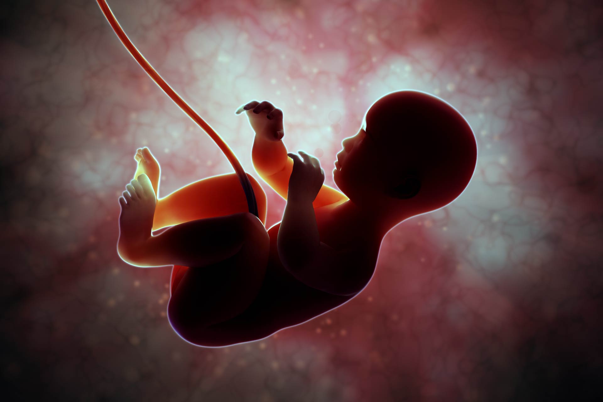
Immune cells populate the fetal developing intestine by week 13 of gestation. At this stage memory T cells are abundant and capable to activate inflammatory responses to foreign antigens, suggesting that specific microbes may influence T cell activity. However, the presence of microbes within the human fetal intestine and their influence on mucosal immunity has not been elucidated. In this study the team investigated on how microbiomes have the potential to influence immunity in the fetal intestine.
Firstly, to determine whether bacteria were present in the intestine in utero, the researchers performed scanning electron (SE) microscopy on fetal terminal ileum specimens from terminated pregnancies (second trimester of gestation). They found evident clusters of tightly packed cellular structures with coccoid bacterial morphology.
To identify and quantify bacteria, using a combination of molecular techniques including 16S rRNA gene burden quantitative PCR and gene sequencing, the team focused on the mid-segment (n = 40) of the small intestine. They identified eighteen taxa significantly enriched in fetal meconium compared to controls (procedural swabs and fetal kidney). In particular:
- The 16S rRNA analysis found two highest fetal meconium taxa, Lactobacillus-meconium (LM), and Micrococcaceae- meconium (MM). Other samples were variably enriched by other bacterial taxa, including taxa within Lactobacillus and Micrococcaceae, Bacteroides, Bifidobacterium and Prevotella.
Furthermore, the researchers analyzed the composition of lamina propria (LP) T cells pairing it with meconium 16S rRNA data. Interestingly, they found that:
- LP samples associated with the T cells surface markers, PLZF+ and CD161+ correlated with meconium characterized by high MM, and MM samples showed significantly higher proportions of PLZF+CD161+ T cells compared to all other samples.
Because fetal intestinal memory T cells expressing PLZF and CD16 have been previously associated with inflammatory response, the team have analyzed epithelial cell layer transcriptomes of samples high in MM.
- The gene-set analysis identified the enrichment of transcripts genes associated with intestinal epithelial stem cells such as LGR5, SOX9, NOTCH1 and NOTCH4, with Toll-like receptor (TLR)-signaling (NFKB2 and TNFSF15), phagolysosome function (NOS2), immune cell chemoattraction (CXCL1-3 and CCL20) and macrophage inhibition (CD200).
Importantly, these results indicate that specific gene expression programs related to immune cell recruitment and regulation are activated in samples high in Micrococcaceae.
The researchers then focused on fetal bacteria isolated from monocyte co-cultures, using full-length 16S rRNA sequencing and the SILVA database.
- The bacteria classified as Micrococcus luteus (Micro 36), exhibited a unique fitness advantage over phylogenetically related strains, under culture conditions that mimicked the fetal intestinal environment including placental steroid hormones and immune cells.
Thus, the team further investigated on Micro 36 discovering that this strain was able to grow under limiting conditions such as high progesterone and β-estradiol, in carbon-limiting medium and low cell density, compared to two reference strains MicroRef1 and MicroRef2.
These data suggest that stringent growth conditions of pregnancy hormones and low nutritional substrate availability, allow only limited growth of specific fetal bacterial thus controlling bacterial burden in the fetal intestine during human gestation.
Because the researchers were able to isolate Micrococcus only when co-cultured with monocytes, they hypothesized that these bacteria were able to survive during gestation solely within phagocytic cells. Therefore, the team have isolated primary human fetal intestinal HLA-DR+ antigen presenting cells (APCs), cleared of intracellular bacteria, and incubated with fetal isolated Micrococcus to promote phagocytosis.
- Gentamicin protection assays followed, revealing that Micro 36 remained viable in APCs at 48 h following antibiotic treatment.
These data indicate a capacity of Micrococcus for prolonged intracellular survival, furthermore, the ability of this fetal strain to persist inside phagocytes offers a potential mechanism of endurance in the fetal intestine.
Further analysis on Micro36 using whole-genome sequencing reported Micro36 exhibited 425 unique genes expression compared to control (MicroRef1), including two sterol carrier proteins and a steroid ketoisomerase, which degrades steroid hormones.
These results might explain why Micro36 is able to grow on placental hormones and to remain viable in phagocytes, under pregnancy limiting growth conditions.
In vitro experiments examined the capacity of fetal M. luteus to induce gene expression changes in primary human fetal intestinal epithelial cells exposed to Micro36.
- Micro36 short-term exposure only partially recapitulated features observed in MM-associated epithelium samples, showing and induction in the expression of TLR6 and its downstream regulator NKFB.
Finally, the researcher assessed the capacity of Micro36 to influence primary fetal HLA-DR+ APC function obtained from spleen.
- Micro36 induced fetal APC production of cytokines associated with maturation of intestinal macrophages and reduced levels of tumor necrosis factor (TNF)-α compared to control strains.
- Moreover, Micro 36 exposure resulted in a significant reduction of IFN-γ production in APC cells as compared to control.
These results indicate that Micro 36 limits inflammatory response, inhibiting IFN-γ production perhaps inducing immunotolerance in fetal intestinal immune cells.
The best methods to address low-burden bacteria in the fetal intestine has not been yet described, however in this study molecular methods coupled with comparative genomics identified few bacterial taxa not attributable to contamination, suggesting that conditions in the fetal gut highly limit bacterial growth. The data presented here offer a potential explanation for M. luteus survival in the fetal intestine, regardless nutrient limitation and pregnancy hormones. Also, these results suggest that fetal intestinal immune cells are capable of activating inflammatory pathways in response to bacteria and that fetal M. luteus may inhibit proinflammatory response in APC cells.
In summary this study shows that viable bacteria are present and highly limited at mid-gestation in the fetal intestine and identifies a potential bacterial mechanism of immune regulation.