
What is already known
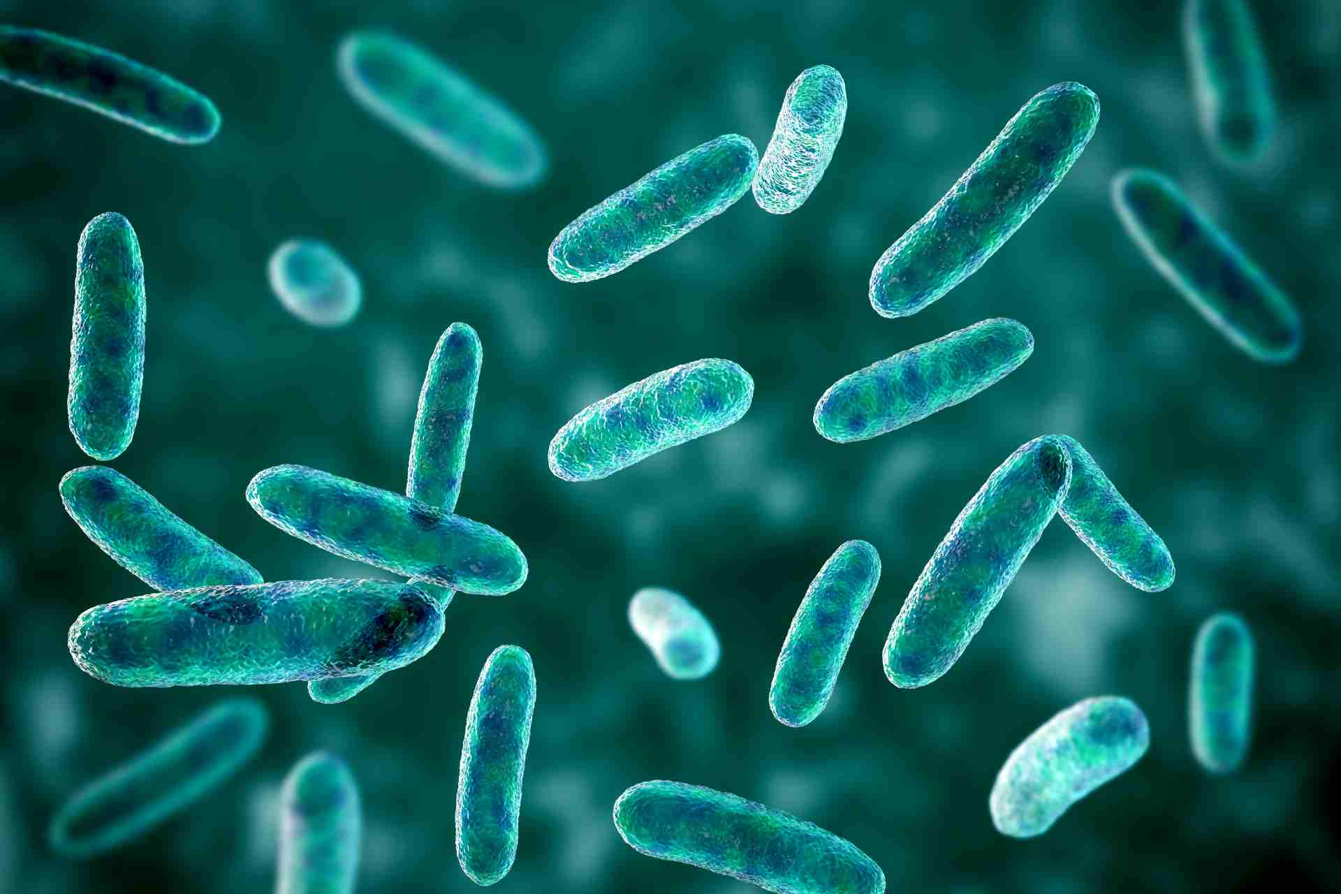
The gut microbiota regulates many body functions, and alterations in its composition have been linked to conditions including cancer, obesity and respiratory diseases. However, there are no standard techniques to image gut bacteria in living animals.
What this research adds
Researchers developed a non-invasive method to image gut bacteria in living mice. The approach is based on the fluorescent labeling of the bacterial cell wall and proved effective in mice with conventional microbiotas as well as in germ-free animals that received a microbiota transplant.
Conclusions
The imaging approach can provide insights into the relationship between mice and their microbiota. It can also offer a glimpse of the cell wall turnover of gut bacteria in living animals.
The gut microbiota regulates many body functions, and alterations in its composition have been linked to conditions including cancer, obesity and respiratory diseases. Now, researchers have developed a method to image gut bacteria in living animals.
The approach, which was detailed in Cell Chemical Biology, can provide insights into the relationship between mice and their microbiota. It can also offer a glimpse of the cell wall turnover of gut bacteria in living animals.
Imaging of animal models is widely used to study biological processes. However, there are no standard techniques to image gut bacteria in living animals. So, Marcos Pires at the University of Virginia and his colleagues set out to develop a method that would allow researchers to tag bacteria with fluorescent molecules and then image them in living mice.
The team set out to target the bacterial cell wall because of its chemical composition: the main component of the bacterial cell wall is peptidoglycan, a polymer composed of two sugars, one of which is linked to short proteins known as stem peptides. One of the characteristics of peptidoglycan is that stem peptides can be tagged with specific amino acids called D-amino acids. Indeed, bacteria are known to swap D-amino acids from the environment into their peptidoglycan scaffold as they grow.
Cell wall tagging
The researchers hypothesized that gut bacteria could be imaged by feeding mice D-amino acid tagged with a fluorescent molecule. So, they used a combination of a modified D-amino acid and fluorophore, a fluorescent chemical compound that can re-emit light upon light excitation.
The fluorescent probe was quickly incorporated into the cell wall of bacteria grown in a dish, the researchers found. Growth media that boosted bacterial growth triggered a rapid loss of fluorescent tagging, likely as a result of faster peptidoglycan remodeling. When bacteria were grown in media promoting slower growth, they showed a slower loss in cellular fluorescence.
“We anticipate that loss of cellular tagging could be a proxy for the kinetics of cellular growth and remodeling and, therefore, may be used to gain insight into [peptidoglycan] remodeling in gut commensal bacteria,” the researchers say.
Illuminating the microbiota
To test the imaging approach in living animals, the researchers fed mice with the modified D-amino acid and the fluorophore, and then imaged the animals two hours later. Mice treated with the fluorescently tagged D-amino acid showed greater fluorescence in their guts compared to controls. The fluorescent signal decreased between four and 22 hours after the last administration of the D-amino acid.
Germ-free mice treated with the fluorescently tagged D-amino acid didn’t have any fluorescence in their guts. But the researchers observed an increase in fluorescence after the mice were given a microbiota transplant, with the cecum and the large intestine — which have a larger load of bacteria — labelling more prominently.
“These results represent a promising first step to potentially extend this strategy in the future into a non-invasive imaging tool that can provide insight into the dynamics of the gut commensal bacteria,” the authors say.